Detecting Durian Leaf Disease (Leaf Blight, Leaf spot, Anthracnose) with Hyperspectral Imaging

The ability to detect durian leaf disease and quantify the percent area of leaf disease at the early stage enables early action with crop management practices, preventing the spread of infection. Common diseases include leaf blight, leaf spot, and anthracnose, which are all caused by the fungus colletotrichum gloeosporioides. They start with young leaves displaying fading symptoms or small light-yellow spots as the fungus grows. With time, the edges of affected parts turn brown with dry and brittle centers. The infection is spread through air and resides in various plants, even in weeds located within durian orchards.
The conventional approach to detect durian leaf disease involves visually checking plants for symptoms or through chemical analysis. These methods can be laborious and ineffective since visible symptoms typically only appear during the middle to late stages of infection. Furthermore, they are time-consuming or necessitate sample extraction that would damage the leaf. Today, non-destructive detection and analysis of durian leaf disease are made possible with the advancement in imaging technologies. The forefront of these technologies is hyperspectral imaging (HSI), a combination of both digital imaging and spectroscopy. With HSI, the plants’ structural and physiological characteristics can be extracted and used to identify diseases before visible symptoms manifest, providing an opportunity to take prompt action and deal with them in time.
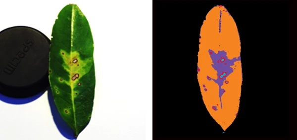
The HSI process entails the capture of spectral and spatial information from plants over a broad range of the electromagnetic spectrum. The data collected, also known as hypercube, are then processed using vegetation indices or algorithms to identify and classify the disease. Below is an example of how the severity of the durian leaf disease can be assessed by classifying, monitoring, and quantifying the infected area on the total leaf area with hyperspectral camera Specim IQ and SpecimINSIGHT software.

Figure 1 – Hyperspectral imaging of durian leaf disease with Specim IQ and SpecimINSIGHT software

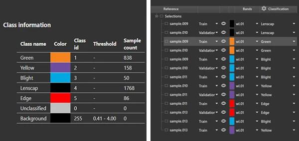
Figure 2 – Class information
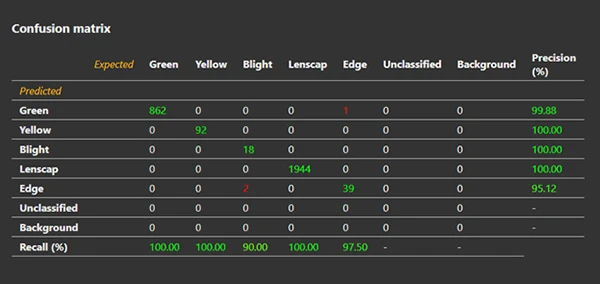
Using Specim IQ and SpecimINSIGHT software, the durian leaf disease can be captured (figure 1) and classified (figure 2). Then, the pixel count can be obtained from the validation sample and applied to the data of confusion matrix (figure 3). From here, we can calculate and characterize the leaf blight and disease sample of durian leaf in quantitative percent area using the pixel count of each classified area.

Figure 3 – Data of confusion matrix
Aside from Specim IQ and SpecimINSIGHT software, Specim, a pioneer in the field of HSI, also offers a board selection of hyperspectral cameras and solutions that are widely used in various plant phenotyping and precision agriculture applications.
Need assistance finding the right hyperspectral camera or solution for your plant phenotyping and precision agriculture applications? Contact our specialists for a free consultation now.
