

Hyperspectral Imaging in Medical Applications – Skin Cancer Diagnosis

Skin cancer is one of the most prevalent forms of cancer around the world. While there are many different types of skin cancer, the three most common are melanoma, Basal Cell Carcinoma (BCC), and Squamous Cell Carcinoma (SCC). BCC and SCC are also sometimes referred to as non-melanoma skin cancer (NMSC). Early detection is vital to the effective treatment of skin cancer. The diagnosis of skin cancer typically involves a preliminary visual examination of the pigmented skin lesions (PSLs) by a dermatologist using the ABCDE (Asymmetrical, Border, Color, Diameter, Evolving) rule before sending for biopsy and histopathological analysis for confirmation. The preliminary examination relies heavily upon the dermatologist’s expertise and can be prone to subjectivity which can lead to unnecessary biopsy and histopathological examination that are invasive, time-consuming, and costly.
Hyperspectral Imaging
In an attempt to reduce diagnostic error and improve efficiency, many are exploring the use of advanced imaging technology in the diagnosis of skin cancer. The forefront of these technologies is hyperspectral imaging (HSI), a combination of spectroscopy and imaging, which can provide non-invasive detection and classification of different skin lesions. HSI captures spectral and spatial information of an object across a wide range of the electromagnetic spectrum. The collected data, also known as hypercube, is then processed (e.g., extract, unmix, classify) to obtain relevant information for the intended applications.
Medical Hyperspectral Imaging (MHSI)
HSI is applied in numerous agriculture and food applications like detecting plant disease and stress, quality inspection of meat, fruits, and vegetables, etc., and is rapidly emerging as a potential tool in various skin cancer studies. For example, Nagaoka et al. (2011) used HSI to develop classification frameworks that can automatically differentiate between melanoma and non-melanoma lesions. Calin et al. (2021) combine HSI with unsupervised anomaly detectors to differentiate BCC and normal skin. Due to its rapid and non-invasive capabilities, HSI has also grown increasingly popular in other medical fields. Miclos et al. (2015) developed an algorithm to map cutaneous tissue oxygen concentration using HSI, while Calin et al. (2015) used HSI to characterize open wounds.
 Specim IQ Hyperspectral Camera
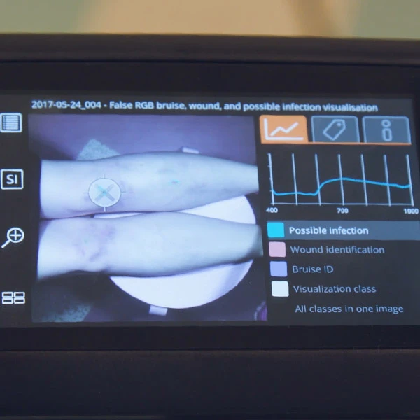
Specim, the leading provider of HSI solutions, offers a wide selection of pushbroom (line scanning) hyperspectral cameras, notably the Specim IQ. A portable visible and near-infrared (VNIR) hyperspectral camera suitable for indoor and outdoor usage, the Specim IQ offers an easy-to-use user interface that allows anyone, whether beginners or experts, to perform HSI measurement and data processing with ease. Unlike most pushbroom hyperspectral cameras, the Specim IQ doesn’t require any external scanner during measurements and operates similarly to a digital camera. Supported by the Specim IQ Studio software that can store and manage HSI data, it allows users to create custom models, applications, and profiles that can be loaded and used in their Specim IQ hyperspectral camera.
Check out this video and see how Specim IQ is used in skin tumor diagnosis.
Wish to learn more about HSI or need assistance finding the right HSI solutions for your applications? Contact our specialists for a free consultation now.