

Introduction to Hyperspectral Imaging Technology

Hyperspectral imaging (HSI) technology has rapidly gained popularity over the past few decades. A combination of spectroscopy and imaging, HSI captures and analyzes images in many different wavelengths across the electromagnetic spectrum. Instead of capturing images using three spectral bands (red, green, and blue) in the visible spectrum like a conventional RGB imaging technology, HSI can capture images in the other wavelengths like infrared, etc., revealing more information about the objects and materials being imaged.
How Does Hyperspectral Imaging Work?
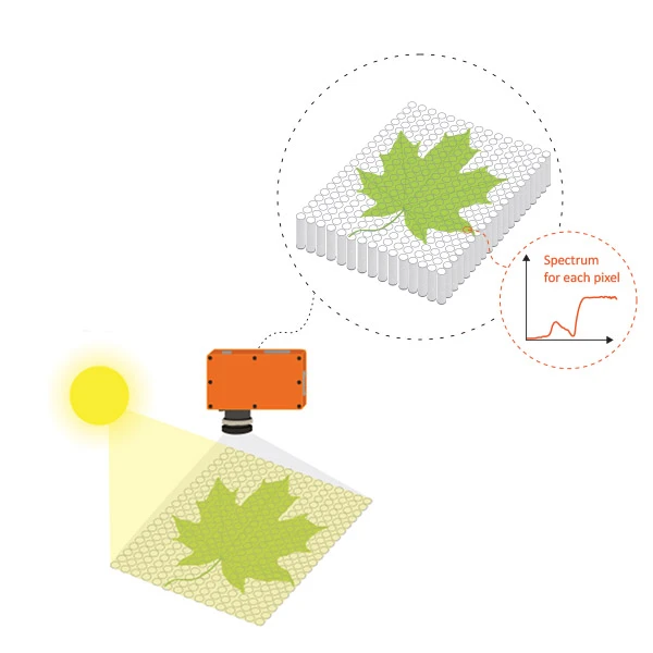
HSI captures images using hundreds and thousands of narrow contiguous spectral bands, providing a detailed spectral signature of the objects in the scene. It involves the use of a hyperspectral camera designed to capture images across a wide range of the electromagnetic spectrum, typically from the visible to near-infrared (VNIR) or shortwave infrared (SWIR) regions.
When the hyperspectral camera captures an image, it records the intensity of light reflected or emitted from each pixel in the image across a wide range of wavelengths. This spectral data is then processed to generate a hyperspectral image, with each pixel containing a spectrum of the reflected or emitted light from the corresponding location on the object. The resulting hyperspectral image can be analyzed using specialized software to identify and differentiate materials based on their unique spectral signatures or to extract valuable information about the composition and characteristics of an object or material.
Hyperspectral Imaging Applications
With its ability to provide a much more detailed and comprehensive view of an object or material in a rapid, non-destructive manner, HSI is widely used across multiple industries, from agriculture and food to healthcare and even art conservation. For example, it can be used to monitor crop health by analyzing the spectral signatures of plants, allowing growers to detect diseases or nutrient deficiencies before they become visible to the naked eye. HSI can also be used for food quality analysis and control, including sorting fruits and vegetables based on ripeness or sugar content, or grading meat products based on marbling and fat content.
In healthcare, HSI has emerged as one of the promising tools for wound care and skin cancer diagnosis. Through analyzing the unique spectral signatures of tissues, information regarding tissue health, like oxygenation, blood circulation efficiency, etc., can be obtained to help monitor wound healing progression or differentiate between healthy skin tissue and cancerous tissue. HSI is also increasingly being used in the field of art conservation. Using HSI, spectral signatures of paintings and other artifacts can be obtained and analyzed to identify the pigments, binding media, etc., used or even reveal details like under-drawings that are invisible to the naked eye.
Specim Hyperspectral Cameras and Solutions
Specim, part of Konica Minolta Group, offers an extensive selection of pushbroom (line scan) hyperspectral cameras and solutions that are widely used in various research and industrial applications. From the compact Specim IQ hyperspectral camera designed to be used in laboratories or on-site operations and industrial-grade Specim FX series hyperspectral cameras that can easily fit into any new or pre-existing machine vision system to airborne hyperspectral systems.

Specim IQ hyperspectral camera (left) and Specim FX10 hyperspectral camera (right).
Image courtesy of SPECIM, SPECTRAL IMAGING LTD.
Interested to find out more about Specim hyperspectral cameras and solutions, or perhaps require assistance finding the right ones for your application needs? Our HSI experts are here to help. Contact us for a free consultation now.